Study approval, registration and safety oversight
This was a single-arm, open-label, phase 1 clinical study designed primarily to evaluate the safety of transplantation of human iPSC-NS/PCs in patients with subacute complete traumatic SCI. Exploratory efficacy outcomes, including neurological and functional recovery, were evaluated as secondary end points.
The study protocol and any subsequent amendments were reviewed to ensure compliance with the Act on the Safety of Regenerative Medicine and approved by the Certified Committee for Regenerative Medicine at Keio University (R2016001) and the Japanese Ministry of Health, Labour and Welfare. The study was registered with the University Hospital Medical Information Network Clinical Trials Registry (UMIN000035074) and in the Japan Registry of Clinical Trials (jRCTa031190228). The long-term observational follow-up was approved by the Ethics Committee of Keio University (approval number 20221199) and registered in the University Hospital Medical Information Network (UMIN000050104). Transplantation was performed at Keio University Hospital (Tokyo, Japan), and subsequent rehabilitation was conducted at Murayama Medical Center (Tokyo, Japan). The subsequent observational study beyond 52 weeks was performed at Keio University Hospital. There was an investigator initiated clinical study and conducted without industry sponsorship. Patients and the public were not involved in the design, conduct, reporting or dissemination plans of this trial.
After transplantation of the first participant, the absence of safety concerns was reviewed at 3 months by an independent data-monitoring committee established for this clinical study, after which transplantation of subsequent participants was initiated.
Participants
Participants with traumatic SCI classified as AIS grade A were eligible. Key inclusion criteria were age 18 years or older, and an NLI between C3/4 and T10. Transplantation was scheduled between 14 and 28 days after injury, and AIS grade A status was reconfirmed on the morning of transplantation. Exclusion criteria included multiple-site SCI, transection injury, or dural rupture on MRI, a history of SCI, or other spinal cord or subarachnoid disease, as well as pre-existing neurological disorders that could independently cause motor impairment. Participants were also excluded if they had clinically significant abnormalities in laboratory test results, severe respiratory failure, major organ damage or other conditions that would preclude rehabilitation, safe administration of immunosuppressants or reliable neurological assessment. Full inclusion and exclusion criteria are described in the study protocol (Supplementary Information) and have been described previously9. Written informed consent was obtained after explanation of study risks, including those associated with first-in-human iPSC-NS/PC transplantation, as well as alternative treatment options.
Sex was recorded based on participant self-report. Sex was not considered in the study design because this phase 1 study enrolled all eligible participants who met the predefined inclusion and exclusion criteria during the study period. Participants did not receive financial compensation for participation. Medical costs related to study-specific procedures and follow-up assessments were covered by the study.
Preparation of human iPSC-NS/PCs
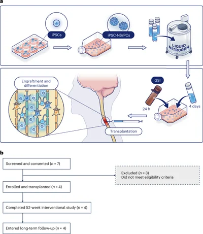
The integration-free human iPSC line YZWJs513 was established under GMP conditions through the iPS Cell Stock Project organized by the Center for iPS Cell Research and Application (Kyoto University). It was created using umbilical-cord blood cells obtained from a healthy volunteer homozygous for most common HLA types in the Japanese population: HLA-A*24:02, HLA-B*52:01, HLA-DRB1*15:02, HLA-C*12:02, HLA-DQB1*06:01 and HLA-DPB1*09:0111. The iPSCs were then differentiated into NS/PCs at Osaka National Hospital using a previously reported protocol for EB-NS/PCs7. In brief, the iPSCs were induced into NS/PCs using serum-free culture of embryoid body-like aggregates with the quick reaggregation (SFEBq) protocol for 14 days, and then expanded using the neurosphere culture technique34,35. The cells from fourth passage were designated as the final cell product.
This final product, designated ONH-iPSNPC-003, met the predefined quality standards. These included characteristics of NS/PCs such as morphology, phenotypic marker expression and terminal neural differentiation capacity, as well as purity (absence of undifferentiated cells), chromosomal normality and absence of genomic mutations associated with reported tumorigenesis risks (Extended Data Table 1 and Extended Data Fig. 1). Quality control testing was performed in compliance with GMP according to validated standard operating procedures established through multiple (three or more) preclinical validation studies, and all results were reviewed by an independent quality assurance unit. The cells were cryopreserved in liquid nitrogen, shipped to Keio University and stored until a potential candidate was identified. At 4 days before transplantation was scheduled to take place, the cells were thawed and cultured to allow them to recover.
Cells were then treated with [N-(3,5-difluorophenacetyl)-l-alanyl]-S-phenylglycine t-butyl ester (DAPT), a γ-secretase inhibitor that is used to suppress Notch signaling3. This treatment was continued for 24 h. Before transplantation, all culture supernatant was carefully removed to ensure that no residual DAPT or culture medium remained. The iPSC-NS/PCs were then resuspended in artificial cerebrospinal fluid for injection.
Confirmation of the long-term stability of the iPSC-NS/PCs
NS/PCs derived from the same iPSC line as those used in the clinical study, but manufactured as a separate batch one year earlier using the same protocol and meeting the same quality standards, were used to evaluate long-term stability. Four predefined parameters were assessed immediately after cryopreservation, at 3 and 6 months, and subsequently at 6-month intervals up to 5 years. The assessments included viable cell count and viability assays; an ATP assay to measure the cell proliferation rate; flow cytometry assays to evaluate the expression of cell surface markers such as PSA-NCAM and GD2 and confirm cell identity; and immunostaining for βIII-tubulin and GFAP to assess differentiation potential and potency. The results are presented in Supplementary Table 2.
Surgical procedure
Before surgery, preoperative MRI was used to identify the rostrocaudal level of the SCI epicenter and to measure the anteroposterior diameter of the spinal cord at that level. The depth corresponding to the injury epicenter was calculated as half of the anteroposterior diameter.
Under general anesthesia, participants were placed prone, and the spinal cord was surgically exposed. Intraoperative ultrasonography was used to identify the injury epicenter and to confirm that its depth and location were consistent with the preoperative MRI findings. A microsyringe was preloaded with the cell suspension containing 2.0 × 10⁶ iPSC-NS/PCs, and the needle depth was preset using a depth stopper according to the preoperatively calculated distance. The needle was manually advanced toward the injury epicenter while avoiding blood vessels. Cell injection was performed slowly over at least 60 s, followed by a 3-min dwell time with the needle left in place before withdrawal. The absence of cell leakage was confirmed, after which the surgical field was irrigated and the wound was closed in a standard manner.
Immunosuppression
Perioperative immunosuppression was provided using tacrolimus. Tacrolimus administration was initiated 1 day before transplantation via intravenous infusion and maintained at a target trough level of 5–10 ng ml−1. Once oral intake became feasible, tacrolimus was switched to oral administration and maintained within the same target trough range for 6 months. Tacrolimus was then gradually tapered and discontinued by 9 months after transplantation. Blood trough levels were regularly monitored to ensure adherence to the target range and to minimize immunosuppression-related AEs.
Mixed lymphocyte reaction assay
An MLR assay was performed to assess donor–recipient immunological compatibility. Peripheral blood (20 ml) was collected from each participant before initiation of the immunosuppressive therapy as the responder cell source. iPSC-NS/PCs used for transplantation were prepared as stimulator cells. At least 3.0 × 10⁶ cells were collected, suspended in culture medium without air exposure, and stored at 4 °C until use. Peripheral blood (10 ml) obtained from an unrelated healthy volunteer, recruited under a separately approved ethical protocol with written informed consent, was used as an allogeneic control. The MLR assay was conducted by an external clinical laboratory (SRL) according to their standardized procedures.
Rehabilitation
In-hospital rehabilitation was provided within the usual therapeutic range, as defined by the Japanese national health insurance system24l
(2024).") during the 52-week interventional study period. That is, for the first 90 days after injury, rehabilitation was provided for up to 3 h per day, and up to 2 h per day thereafter. Rehabilitation included physical therapy, occupational therapy and dysphagia rehabilitation, all of which were tailored to each participant’s neurological status. No advanced interventions, such as electrical stimulation or robotic-assisted therapy were employed.
Outcome measures
During the 52-week interventional study period, safety was the primary outcome and was assessed through systematic monitoring of all AEs. Exploratory efficacy outcomes were evaluated using validated neurological, functional, and patient-reported outcome measures.
Neurological function was assessed using the ISNCSCI10, including motor and sensory scores and neurological severity was additionally categorized using the modified Frankel classification36,37. Functional independence was evaluated using SCIM III12. Neuropathic pain was assessed using the PainDETECT questionnaire15 and the NPSI16 and spasticity was evaluated using the MAS17. These outcome measures were assessed at 1, 3, 5 and 8 weeks after transplantation and subsequently at 4-week intervals until 52 weeks after transplantation.
In the subsequent long-term observational follow-up, neurological and functional outcomes were assessed using ISNCSCI motor and sensory scores and SCIM III, and pain and spasticity were evaluated using PainDETECT and MAS. These assessments were performed at 6-month intervals for the first 3 years after transplantation and annually thereafter.
MRI acquisition and evaluation
MRI of the cervical spine was performed at baseline before cell transplantation, at predefined time points during follow-up, including at 3, 12, 24 and 52 weeks after cell transplantation during the interventional study. During long-term observational follow-up, MRI was performed at 6-month intervals for the first 3 years and annually thereafter.
MRI was performed using either a 1.5T MRI scanner (Signa HDxt and Signa Artist, GE Healthcare) or a 3T MRI scanner (Discovery MR750 and Signa Pioneer, GE Healthcare), with a standard cervical spine coil. The imaging protocol included sagittal and axial T1-weighted and T2-weighted sequences. Sagittal images were acquired with slices oriented parallel to the long axis of the cervical spinal cord, and axial images were obtained at cervical levels C1 through C7 with slices oriented parallel to the intervertebral disks. For T1-weighted imaging, repetition time (TR) ranged from approximately 350 to 660 ms and echo time (TE) from 6 to 13 ms. For T2-weighted imaging, TR ranged from approximately 3,100 to 5,000 ms and TE from 80 to 130 ms. The field of view ranged from approximately 230 to 400 mm for sagittal images and 160 to 200 mm for axial images. The flip angle was 90° for all sequences. Slice thickness ranged from 3 to 4 mm for sagittal images and from 4 to 5 mm for axial images.
Contrast-enhanced MRI was not included in the imaging protocol, as contrast enhancement is known to be variable and nonspecific in intramedullary spinal cord tumors, including gliomas, and may also be observed in non-neoplastic conditions such as SCI itself. Accordingly, tumor surveillance was based on serial noncontrast MRI to evaluate temporal changes in spinal cord morphology and signal characteristics around the transplantation site.
MRI evaluation for tumor formation was based on established clinical and radiological features reported for intramedullary spinal cord tumors and related pathological processes38,39,40,41. Intramedullary tumors are generally recognized on MRI as either mass-forming lesions that cause focal spinal cord compression or infiltrative lesions that diffusely involve the spinal cord parenchyma. Accordingly, MRI surveillance focused on identifying both patterns. For mass-forming lesions, tumor formation was defined as the appearance of a newly developed, well-demarcated lesion that was not present on preceding MRI examinations. For infiltrative lesions, particular attention was paid to progressive or heterogeneous signal abnormalities on T1- and T2-weighted images, lesion expansion, ill-defined lesion margins, craniocaudal spinal cord edema, inflammatory signal changes adjacent to the transplantation site and spinal cord swelling, including features described for gliomas, which represent a major category of infiltrative spinal cord tumors40.
All MRI scans were reviewed descriptively by experienced spine surgeons and neuroradiologists, focusing on potential tumor formation and other graft-related pathological changes following iPSC-NS/PC transplantation.
PET acquisition and evaluation
18F-FDG PET imaging of the whole body was performed 24 weeks after cell transplantation to evaluate metabolic activity. Patients were instructed to fast for at least 6 h before tracer injection. A dose of 4.0 MBq kg−1 FDG was administered intravenously, followed by a 60-min uptake period while the patient was in a resting position. Imaging was performed using a PET–CT scanner (Biograph Vision 600, Siemens Medical Solutions), with acquisition covering the entire body from the top of the head to the soles of the feet. A low-dose CT scan was acquired simultaneously for attenuation correction and anatomical localization.
Axial PET images of the cervical spine were reconstructed using standard algorithms. To measure the maximum standardized uptake value (SUVmax), volumes of interest (VOIs) were manually defined to encompass the entire spinal cord cross-section at the axial slice corresponding to the level of transplantation. The SUVmax within each VOI was calculated using dedicated image analysis software (syngo.via, Siemens Healthineers). The SUVmax values were compared to reported physiological uptake levels of the spinal cord in healthy individuals to assess abnormal metabolic activity, with cervical spinal cord SUVmax values ranging from approximately 1.8 to 2.5, as reported in previous studies42,43.
Adverse event assessment
All reported AEs were coded using the Medical Dictionary for Regulatory Activities (MedDRA), v.27.1, to ensure standardized classification across categories. The severity of AEs was graded in accordance with the Japanese governmental criteria for classification of adverse drug reactions, as defined by a notification of the Pharmaceutical Safety Bureau of the Ministry of Health and Welfare (notification no. 80, 1992).
For symptoms considered intrinsic to the underlying SCI, reflecting pre-existing neurological or autonomic dysfunction rather than effects of the intervention, severity grading was performed using the same three-grade classification framework based on a study-specific AE grading reference sheet. This reference sheet was predefined before trial initiation through consultation with an independent data-monitoring committee established for this clinical study, to ensure consistent and appropriate grading in the context of SCI.
According to these criteria, AEs were classified as grade 1 (mild adverse reactions), grade 2 (moderate adverse reactions that were neither mild nor severe) or grade 3 (severe adverse reactions), defined as events that, depending on patient characteristics or clinical circumstances at onset, could potentially result in death or permanent functional impairment that interferes with activities of daily living.
Statistical analysis
All patients who received study treatment were included in the safety and efficacy analysis populations. Continuous variables were summarized descriptively, and categorical variables were presented as counts and percentages. Safety data were summarized descriptively, with point estimates of proportions calculated without confidence intervals. Given the small sample size of this study, no formal hypothesis testing or confidence interval estimation was performed for efficacy end points. Longitudinal changes in motor scores through 52 weeks were summarized descriptively. All enrolled participants were male; therefore, no sex-based analyses were performed. All analyses were performed using SAS v.9.4 (SAS Institute) and R version 4.5.1 (R Foundation for Statistical Computing).
Historical cohort analysis
An exploratory comparison was conducted using data from the JSSCI-DB13,14, a registry of patients with traumatic SCI at the Spinal Injuries Center (Fukuoka, Japan), which included 986 patients registered between 2013 and 2024. From this population, patients classified as AIS grade A at 2 weeks after injury with neurological injury levels between C4 and C8 were identified as a historical cohort for comparison with the present clinical study. Additional eligibility criteria were applied using available registry variables to approximate the inclusion and exclusion criteria of the present study, including exclusion of patients with severe respiratory impairment and those with major comorbidities such as diabetes mellitus, cerebrovascular disease, renal dysfunction, or psychiatric disorders. Patients without available total motor score data beyond 4 weeks post-injury were excluded. The final analysis set comprised 52 patients.
Total motor score at 2 weeks after injury was defined as baseline, and changes from this baseline were evaluated over time using available-case and LOCF approaches. When measurements at a given time point were missing, values at hospital discharge were used if temporally closest. Neurological outcomes were summarized descriptively as changes in total motor score over time and visualized using longitudinal box-and-whisker plots through 52 weeks after injury. AIS grade conversion to C or higher at 52 weeks was also evaluated. No formal statistical testing was performed.
Information on MRI-based exclusion criteria (for example, multilevel SCI, complete transection or dural injury) and laboratory-based exclusion criteria were not available in the registry and could not be applied. Therefore, residual differences in patient selection between the registry cohort and the present study cannot be excluded.
Reporting summary
Further information on research design is available in the Nature Portfolio Reporting Summary linked to this article.