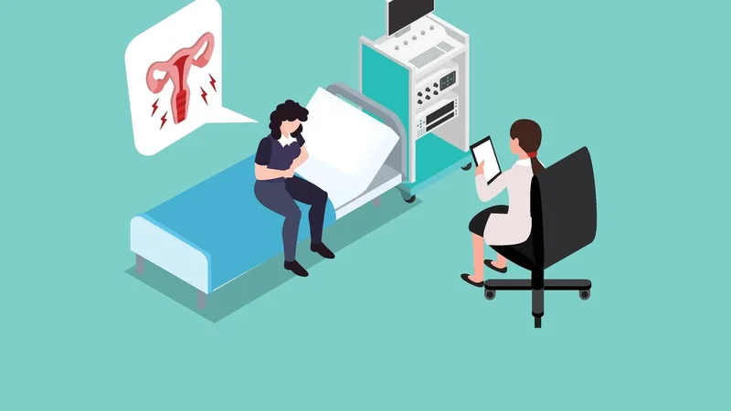
A team of doctors in a private hospital in Bengaluru performed a complex laparoscopic surgery to remove a massive 4.65-kg uterine fibroid from a 28-year-old unmarried woman.
The tumour, which weighed more than a full-term newborn, was removed using 5 mm laparoscopic incisions and a specialised mini-laparotomy morcellation technique. As the fibroid was too large to be extracted through the laparoscopic ports, the team cut it into smaller pieces and removed it through a small 4-cm incision. Despite the extreme size and high-risk location of the mass, doctors at Motherhood Hospitals, where the surgery was done, successfully reconstructed the patient’s uterus.
Undetected for years
The woman from coastal Karnataka had lived with the mass largely undetected for years. She had experienced regular, pain-free menstrual cycles with no heavy bleeding since the age of 15. It was only when she developed abdominal pain around four months before the surgery that scans revealed the large tumour. Pre-operative evaluations revealed that the mass had developed into a rare parasitic fibroid, with additional blood supply from the omentum, the fatty tissue that covers the abdominal organs. Located on the lateral wall of the uterus and close to major pelvic blood vessels and the ureter, the tumour posed an unusually high risk of significant surgical bleeding.
Fibroids in this location are recognised as being among the more surgically challenging to remove. Published data on lateral and broad-ligament fibroids report complication rates of nearly 19% — more than twice those associated with fibroids in other locations. This is largely due to their proximity to the ureter and major uterine blood vessels.
Stabilised for six weeks
The doctors — Madhuri Vidyashankar P., consultant hysteroscopic and laparoscopic surgeon and gynaecologist, and Vijaylakshmi, consultant in obstetrics and gynaecology — and the team spent six weeks stabilising the patient’s elevated thyroid levels and optimising her haemoglobin before identifying and ligating the blood vessels supplying the fibroid. The team then separated the mass from the uterus, reconstructed the uterine wall and preserved the uterus. The patient is recovering well and has reported significant relief, hospital authorities said.
“This was a technically demanding surgery because we were dealing with an exceptionally large fibroid in a very restricted space, located close to major pelvic blood vessels and the ureter. The fibroid had also developed an additional blood supply from the omentum, making control of the feeding vessels critical before we could safely remove it. Once the blood supply was controlled, our priority was to separate the fibroid carefully, reconstruct the uterine wall and preserve the uterus,” said Dr. Vidyashankar.
Complex gynaecological problems
Beyond the clinical success of this procedure, the case points to a serious trend of complex gynaecological problems becoming more common among young women in their thirties, which was not common before among this demographic bracket.
The shift is borne out in global data too. In the most recent ‘Global Burden of Disease’ study, India’s fibroid incidence has been seen rising faster than in almost any other region studied worldwide, with new diagnoses increasingly appearing from adolescence onward rather than only in mid-life.
This emphasises the urgent need for regular preventive check-ups for young women. Routine pelvic ultrasounds during annual health exams are essential for catching hidden growths early, allowing for timely medical management before a condition threatens organ safety, overall health, or future fertility, the doctor added.
Published - August 22, 2026 09:09 pm IST