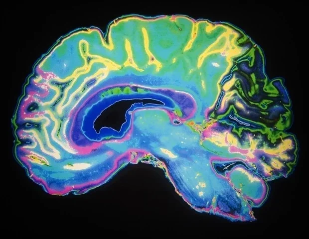
Nanoplastics-plastic particles smaller than one micrometer-are generated through the degradation and abrasion of plastic products and are increasingly detected in food, drinking water, air, and biological tissues. Although the presence of plastic particles in animals and humans is well-established, determining how nanoplastics are distributed within complex organs, such as the brain, remains challenging.Now, a research team from the National Institute for Environmental Studies (NIES), The University of Osaka, and Waseda University has established a workflow to visualize the three‑dimensional distribution of nanoplastics across the neonatal mouse brain without tissue sectioning. The researchers focused on the neonatal period, characterized by rapid brain growth and immature biological barrier systems. Conventional approaches generally rely on tissue sections, allowing only two‑dimensional observation and making it difficult to capture whole‑brain distribution patterns or compare signal intensities across different brain regions. To overcome these limitations, the team combined a tissue-clearing technique with light-sheet fluorescence microscopy, enabling three-dimensional imaging of the brain.
Size‑dependent biodistribution of nanoplastics in neonatal mice
Fluorescently labeled polystyrene nanoplastics were orally administered to neonatal mice, and tissues-including the brain-were collected 24 hours later for imaging. The analyses revealed a clear particle size dependence in biodistribution, with greater accumulation of nanoplastics with a diameter of 50 nm than of nanoplastics exceeding 500 nm in several organs, including the intestine, kidneys, and brain. Signals from 500 nm particles were weak across all examined organs. This study focuses on methodological development and visualization capabilities and does not directly assess environmentally relevant exposure levels or potential health risks.
Whole-brain three-dimensional visualization using tissue clearing and light-sheet fluorescence microscopy
To enable detailed analyses of nanoplastic distributions, the researchers applied a tissue-clearing technique (SeeDB2G) to neonatal mouse brains. This approach rendered the brain optically transparent, allowing fluorescence imaging of deep brain regions without physical sectioning. Subsequent imaging using light-sheet fluorescence microscopy enabled three-dimensional visualization of the nanoplastic distribution throughout the whole brain.
Quantitative analysis of the region-specific distribution in the brain
Whole‑brain three‑dimensional imaging showed that 50 nm nanoplastics were widely distributed throughout the neonatal brain. Quantitative analysis further demonstrated relatively higher fluorescence signals in the thalamus and brainstem than in other regions, such as the cerebral cortex and cerebellum. These regions are located near the ventricular system, suggesting a potential association with cerebrospinal fluid circulation or developmental characteristics of barrier systems in early development. It should be noted that this study is based on relative comparisons of fluorescence intensity and does not directly quantify the absolute amount of nanoplastics or identify specific invasion pathways. The elevated signals observed in the thalamus and brainstem may reflect particle entry processes or circulation and clearance dynamics, and further detailed analyses are required to clarify these mechanisms.
Validation of fluorescence signals
Because fluorescence‑based detection can be affected by dye leakage or tissue autofluorescence, the researchers conducted a hyperspectral imaging analysis to validate the signals. The fluorescence signals detected in both cells and intestinal tissues exhibited spectral characteristics consistent with those of polystyrene nanoplastic, confirming that they were particle-derived rather than arising from free dye or tissue autofluorescence. Confirmation through multiple independent analytical approaches supports the reliability and robustness of the observed distribution patterns.
This study demonstrates that nanoplastics can accumulate in the brain and other organs during the neonatal period and that this distribution can be visualized three-dimensionally at the whole-brain level. The newly established method provides a foundational analytical platform for investigating the biodistribution and spatial localization of nanoplastics. The study does not directly evaluate toxicity or health effects, nor does it reflect environmentally relevant exposure levels. However, it offers a robust framework for future studies aimed at elucidating invasion pathways, circulation dynamics, and organ-specific distribution patterns of nanoplastics.
The findings are scheduled for publication in the Journal of Hazardous Materials Advances on June 29, 2026, under the title "Whole‑Tissue Distribution Analysis for Visualization of Nanoplastics in the Neonatal Mouse Brain."
Source:
Journal reference:
Mi, Y., et al. (2026) Whole-tissue distribution analysis for visualization of nanoplastics in the neonatal mouse brain. (2026). Journal of Hazardous Materials Advances. DOI: 10.1016/j.hazadv.2026.101353. https://www.sciencedirect.com/science/article/pii/S2772416626003499?via%3Dihub